


 68 / 80

Practitioner's Corner
J Investig Allergol Clin Immunol 2019; Vol. 29(5): 378-398
© 2019 Esmon Publicidad
consolidation, areas of ground-glass opacity, micronodules,
and tree-in-bud opacities. Esophagogastroscopy showed a
dilated esophagus. Esophageal high-resolution manometry
revealed a hypertensive lower esophageal sphincter that did not
relax on swallowing; similarly, there was no peristaltic wave
in the esophagus. The patient was diagnosed with achalasia.
When the test results had been collected and the medical
history was being completed, the patient reported that for
at least 3 years, she had experienced nocturnal vomiting
containing undigested food; her nasal discharge had also
contained food particles. The symptoms were associated with
persistent cough.
Once the diagnosis of achalasia was confirmed, the patient
was referred for surgery (peroral endoscopic myotomy). A
check-up 2 months after surgery revealed that cough, dyspnea,
rhinorrhea, and nocturnal vomiting had considerably abated.
All asthmatic medications were discontinued. Spirometry
results returned to normal values (FEV
1
, 3.89 L [121%]; FVC,
4.3 L [117%]; FEV
1
/FVC, 90%). The result of the methacholine
challenge test performed at that time was negative.
The present case concerns a patient with respiratory
symptoms resulting from achalasia that were misdiagnosed as
severe asthma. In fact, the symptoms reported were caused by
recurrent aspiration of small amounts of gastric content that
occurred largely at night over a period of a few years. The chest
CT scans performed on admission to the Allergy Department
were characteristic of bronchiolitis and reflected chronic
bronchiolocentric inflammation caused by recurrent aspiration.
The clinical picture and imaging scans pointed to a diagnosis
of diffuse aspiration bronchiolitis (DAB) complicated by
incidents of aspiration pneumonia. The term diffuse aspiration
bronchiolitis was first used by Matsuse et al [4] as a name
for a chronic inflammation of the bronchioles produced by
frequent aspiration of foreign particles. Although DAB is
usually diagnosed in the elderly, it has been reported in younger
patients, with clinical manifestations similar to those found in
the elderly [5-8]. In younger patients, the major risk factors
responsible for DAB are dysphagia due to achalasia and
gastroesophageal reflux disease with concomitant recurrent
aspiration.
In the case we report, the diagnostic delay may have
been caused by various factors. First, apart from a 5-year
history of vomiting that was erroneously interpreted as a
consequence rather than the cause of coughing, there were no
accompanying symptoms characteristic of achalasia. Second,
auscultatory phenomena were interpreted as asthmatic
wheezing, while they might in fact have resulted from
pressure on the trachea and/or bronchiolitis, which can also be
responsible for variations in airflow obstruction in spirometry.
Third, achalasia is a rare disorder, diagnosed mostly in elderly
adults (generally during the sixth decade of life, with an
estimated prevalence and incidence, respectively, of 10.82
cases per 100 000 and 1.63 cases per 100 000 [9]). Fourth, the
primary symptoms of achalasia are mostly gastrointestinal,
whereas respiratory symptoms are less frequent. In up
to 40% of cases of achalasia, pulmonary disorders such
as cough, wheezing, and recurrent aspiration can occur,
although DAB is very rare [10]. Bronchiolitis associated
with chronic aspiration can considerably hamper diagnosis.
DAB should be considered in patients with respiratory
symptoms such as chronic cough, wheezing, obstruction,
persistent radiologic abnormalities in high-resolution CT, and
a high risk of aspiration. Given the scope of the respiratory
changes we report, the possible consequences of a further
delay in surgical treatment of achalasia could be serious.
Our findings confirm the prevailing stance of asthma experts
who claim that if asthma symptoms persist despite intensive
pharmacological treatment, it is advisable to revisit the
patient’s clinical history, bearing in mind the possibility of
a diagnosis that mimics asthma.
Funding
The authors declare that no funding was received for the
present study.
Conflicts of Interest
The authors declare that they have no conflicts of interest.
References
1. Chung KF, Wenzel SE, Brozek JL, Bush A, Castro M, Sterk PJ,
et al. International ERS/ATS guidelines on definition, evaluation
and treatment of severe asthma. Eur Respir J. 2014;43:343-73.
2. Kim S, Lee CH, Jin KN, Cho SH, Kang HR. Severe asthma
phenotypes classified by site of airway involvement and
remodeling via chest CT scan. J Investig Allergol Clin Immunol.
2018;28(5):312-20.
3. Robinson DS, Campbell DA, Durham SR, Pfeffer J, Barnes PJ,
Chung KF. Systematic assessment of difficult-to-treat asthma.
Eur Respir J. 2003;22:478-83.
4. Matsuse T, Oka T, Kida K, Fukuchi Y. Importance of diffuse
aspiration bronchiolitis caused by chronic occult aspiration in
the elderly. Chest. 1996;110:1289-93.
5. Akritidis N, Gousis C, Dimos G, Paparounas K. Fever, cough,
and bilateral lung infiltrates: achalasia associated with
aspiration pneumonia. Chest. 2003;123:608-12.
6. Matsuse T, Teramoto S, Matsui H, Ouchi Y, Fukuchi Y.
Widespread occurrence of diffuse aspiration bronchiolitis
in patients with dysphagia, irrespective of age. Chest.
1998;114(1):350-1.
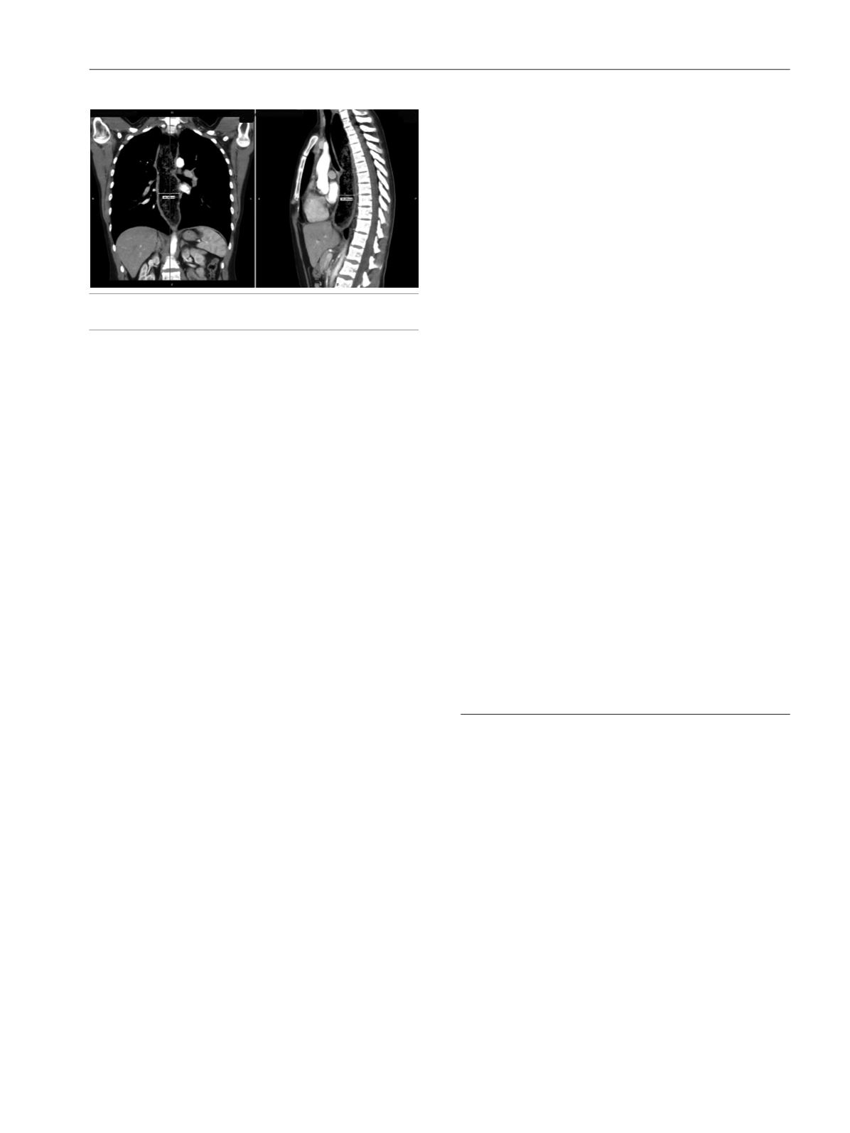
Figure.
Computed tomography scan showing massive dilatation of the
esophagus and tracheal compression.
395